A New Machine Learning Method and System For Automated Disease Detection from Chest Radiography Images
Description: An important game changer in the recent times is the application of machine learning techniques in healthcare domain. It is no wonder that the current research on computer analysis of medical images has achieved a substantial importance with a rapid technical progress in artificial intelligence and machine learning areas. In this work, we aim to propose an explainable system and method to identify near similar medical images by leveraging linear algebra so as to discriminate between normal, pneumonia and COVID-19 chest radiograph images. Decisions made by applying machine learning models which are built by leveraging linear algebra shall have strong mathematical basis. Also, such an explainable machine learning based medical imaging system that combines machine learning and mathematical techniques for accurate disease diagnosis promotes transparency, clear, interpretable diagnostic reports, ultimately assisting healthcare professionals in taking informed decisions. The experiment results have proved that the proposed method for disease diagnosis from chest radiography images discriminates effectively between normal, pneumonia and COVID-19 images.
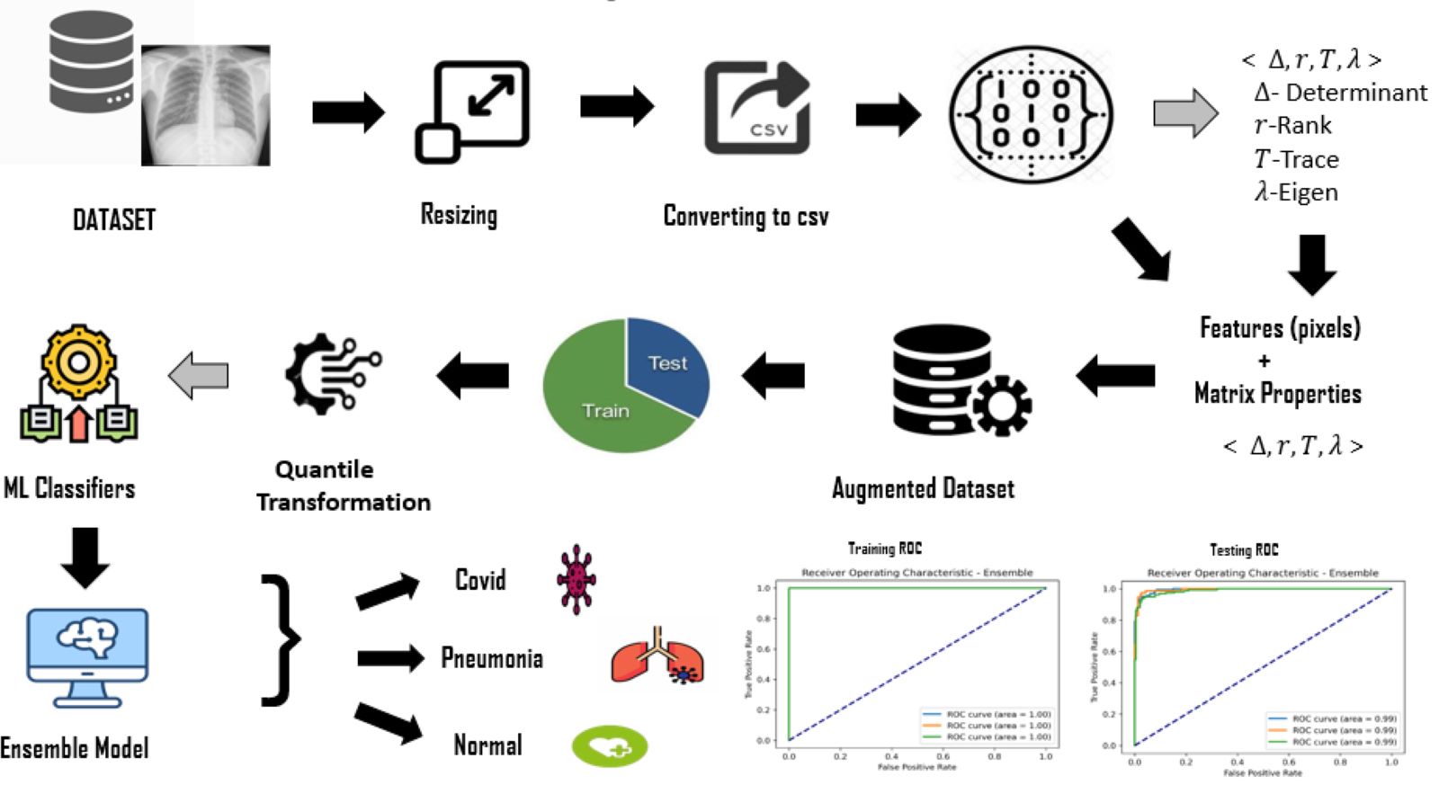
The major challenge in disease diagnosis using medical images for explainable machine learning lies in identifying appropriate and contributing image pixel features which can help the learning machines to perform better thereby achieving high detection rates. Feature engineering is thus an important step in building a good machine learning model. The idea is to build an explainable AI model, yet computationally efficient and whose learning process is guided by proposed feature engineering phase. The output of the proposed method is explainable to correlate the diagnostic decision. Such an output by learning machines can aid radiologists in decision making. The proposed idea is to use an Ensemble model that fits on the pixel features and extracted matrix properties drawn out by the feature extraction phase. The chest radiography images are pre-processed by resizing into same size. The matrix properties are extracted from these images and are augmented to the data after flattening the X-ray image. After augmenting the extracted features to original pixel features, the combine features are used to train the machine learning classifiers. The ensemble model is then fit to create a balance among detection rate, specificity, and balanced accuracy of the final model.
Organisation: VNR Vignana Jyothi Institute of Engineering and Technology
Innovator(s): V. Sravan kiran, R. Adhi Venkata Pavan, Dr. Rajesh Appusamy, Dr. V. Radhakrishna
Category: Information Technology, AI and ML
Country: India